afib with ST elevation
First Glance:
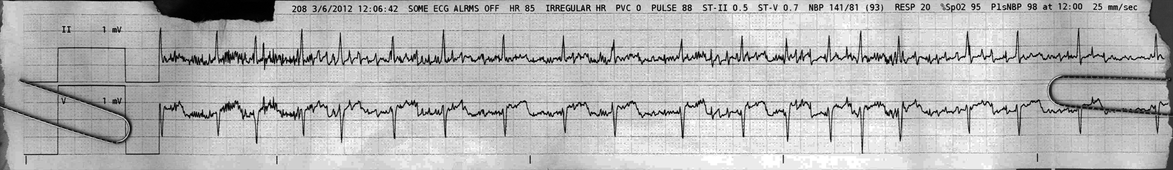
From across the room it looks like a brisk irregularly irregular rhythm with artifact, with some ST elevation in the chest lead.
Discussion:
There is a lot of fine choppy baseline artifact, but the irregularly irregular narrow-complex rhythm and the lack of any obvious identifiable P waves in either lead (including the chest lead at 200%) suggest atrial fibrillation. The choppy fluttery look of the baseline between beats 4 and 6, and again later in the strip might be artifact, or it might be bits of atrial fibrillation with larger reentry pathways or bits of flutter in one atrium. You can convince yourself there might be P waves with normal PR intervals before some beats, but there is no consistency and it’s hard to make a good argument for a specific rhythm from this strip. It’s likely afib, but we can’t be 100% sure there isn’t an underlying regular rhythm with some ectopy.
We see 1.5 to 2mm of ST elevation in the chest lead. This is a soft call, as tele often gets the ST elevation wrong compared to a real 12L, but it doesn’t hurt to notice. The gain is 200% so this is only 1mm of ST elevation, but still possibly significant. The rate is around 100 bpm.
Here’s a strip from the same patient, two hours earlier:
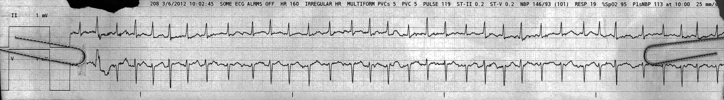
Here we see a faster rhythm, completely irregularly irregular- consistent with afib. There is nothing much that looks like a tempting P wave here. If we were to diagnose by the company it keeps we might say that if this is afib, so was the first strip, but it doesn’t work like that so we still can’t be definitive about the initial strip you saw.
Beat 2 looks like a fusion beat, with a similar morphology in II but with a PVC-like look in the chest lead. Notably, this strip does not show the ST elevations we saw two hours later, despite a significantly faster rate. While I might make much of 1mm of ST elevation in a tele strip, if it wasn’t there beforehand than I am more concerned.
Final Impression?
(note that the first strip shown is the later one chronologically)
1) Likely atrial fibrillation @ ~85 bpm, can’t rule out other irregular SVT.
2) afib with RVR @ ~150 bpm, possible new ischemia/injury
Management implications:
To make a firm diagnosis of the first lead reduce artifact (you want a warm, calm patient, a good lead, or a 12L). Thoughtful rate control will be important in this patient.
A 12l will be necessary to evaluate properly for ischemia/infarction.
The Take-home Point:
Fibrillation is the existence of many many tiny reentry pathways in the myocardium. Atrial fibrillation might have dozens of tiny reentry pathways, all overlapping electrically, so the baseline generally looks fine and unpredictable. Atrial flutter is another atrial reentry rhythm, but there is only one single reentry pathway, so we see the one large regular flutter baseline. If there is something in between with only a few simultaneous reentry pathways, or if there is one or two large dominant reentry pathways, or if one of those phenomenae are intermittent, perhaps it could cause an irregular but flutter-esque baseline such as we see here. The term ‘fib-flutter’ is thrown around a lot by savvier folks than I. By my most recent EP tutorial it is defined as alternating between flutter and fib (a common occurrence).
![]() Two star strip. The meat and potatoes.
Two star strip. The meat and potatoes.