atrial flutter
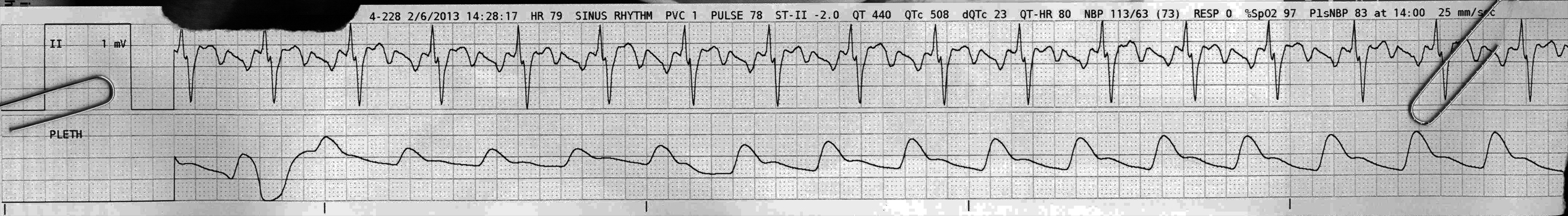
First Glance:
From across the room it looks like atrial flutter
Discussion:
The gain on this lead has been jacked up to 200% and a lead has been selected that shows obvious flutter waves, so these F waves are all but slapping you in the face. This looks like flutter with a 3:1 block (an unusual combination).
This is lead II, and the flutter waves are deeply negative.
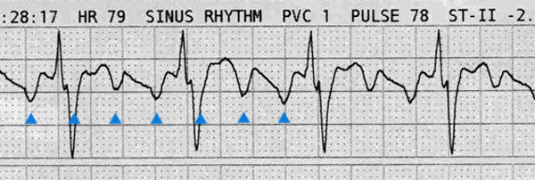
How do I say negative? Well, the sharpest deflections are negative. You could easily imagine that the baseline is actually composed of wider positive deflections, but the pros tell me to pick the ‘sharper’ deflection and that is what makes intuitive visual sense.
With deeply negative F waves in the inferior leads, this is probably typical atrial flutter which classically runs at ~300 bpm +/- 20 or so. Positive F waves in II would be more likely to be type 2 flutter, which is associated with significantly faster flutter rates. However, if you look closely there are six small boxes between F waves, which means the flutter rate is 1500 / 6 = 250 bpm. This is on the slow side for typical flutter, and on the high side for atrial tachycardia.
How could we differentiate? Atrial flutter classically has a ‘sawtooth’ baseline, which we see nicely here. This is to say that the baseline never really flattens out between F waves, where we would expect flat baseline in ectopic AT.
Is that an unusual axis on the QRS wave? Wouldn’t we expect it to be predominantly positive?
Yes. The deep S in the QRS is a result of the superimposed deeply negative F wave. The actual QRS probably has a completely normal axis.
Final Impression?
typical atrial flutter with 3:1 block, for a ventricular rate of around 80 bpm.
Management implications:
Examine some other leads to see the flutter waves. If there are sections of flat baseline between the P waves this would push us back towards an ectopic atrial tachycardia. The differentiation of the two might be largely theoretical, as the large reentry circuits that generally cause atrial flutter can look electrically identical to the rapid P waves of an excited ectopic atrial focus (or a focal micro-reentry circuit). We make our guesses, but in the end keep the differential on the backburner as the treatment for flutter and ectopic atrial tach are quite different, depending on the clinical situation.
The Take-home Point:
The rate is a clue, and comes with a caveat: Yes, classic atrial flutter is 150 bpm- a 300 bpm flutter rate with a 2:1 block, but we’ve seen 3:1 block here, and there is plenty of 4:1 block, and there can be higher blocks still, so all kinds of rates could be possible. Further, while ‘typical’ flutter generally has a flutter rate of 280-320 bpm, atypical flutter can be as fast as 400 bpm, so now if you include all the block possibilties you could really see any possible rate with atrial flutter. Making this diagnosis will boil down to see regular flutter waves marching across the strip at a rate that is higher than expected for atrial tach with a sawtooth baseline.
![]() Two star strip. The meat and potatoes.
Two star strip. The meat and potatoes.