wide complex rhythm
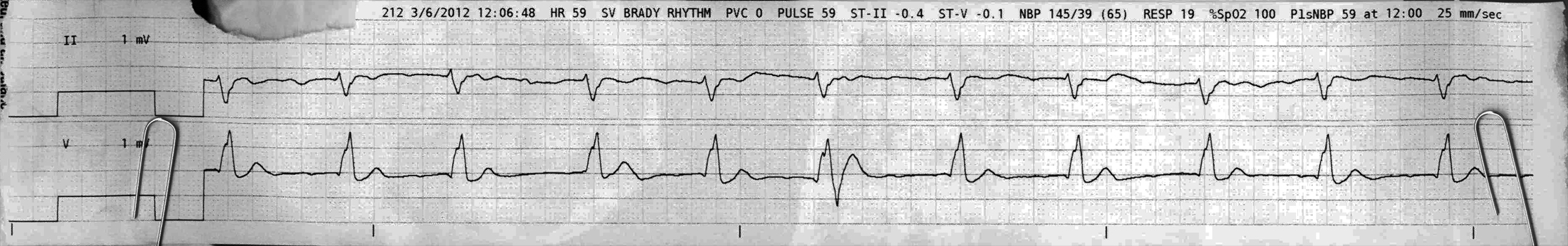
First Glance:
From across the room it looks like a regular wide complex rhythm at a friendly rate.
Discussion:
We see here a fairly regular rhythm @ ~65 bpm with a little RR variability, but not enough to obviously be atrial fibrillation. Afib can look exactly this regular sometimes, but if you watch more tele you’ll see it get choppier. This one stayed just like this.
If you look closely, you can pick out subtle deflections that look like P waves- best seen in the chest lead. There are a bunch with a PR interval of around 240 ms (marked with red stars below). There are also some other deflections that might be P waves, but they are not seen as much, and don’t seem as consistent (marked with blue triangles).
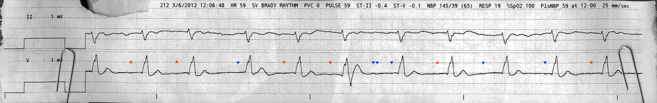
These deflections are subtle enough that perhaps there are P waves with a 240ms PR interval before more QRS complexes but we just don’t see them. Perhaps they are artifact. Let’s look a little closer.
Beats 2 and 3 have this maybe-sinus P before them. Then beat 4 doesn’t have the expect P wave, and the interval is longer. There is a shallow deflection with a shorter interval before the beat 3 QRS, but it isn’t as convincing. One way to get this would be two sinus P waves, followed by a sinus pause and a junctional escape beat. The delay from the beat 3 P wave to the beat 4 QRS would be a rate of 44bpm, which is consistent with junctional escape. The next two beats also look like the sinus with long PR, followed by another long RR interval and no obvious P wave. Is this another sinus pause with junctional escape?
Or maybe, there is actually a rapid supraventricular rhythm- maybe flutter or AT at a brisk rate with a variable block. That could give you a similar look, and would explain the tendency to be regular but with some variations, ie: if this was atrial flutter with variable 4:1 and 5:1 block.
The rest of the strip has a smattering of maybe-Ps, but the truth is we aren’t going to solve this one definitively from this strip. As much as you want to analyze, we really need more strip (and preferably better strip) to crack this case.
Anything else?
Lead II has a flattened biphasic T, and the chest lead has a bit of ST scooping that looks like digoxin effect. You usually see that in the lateral leads, and if this V lead is a lateral lead that might suggest a LBBB. However, we would not expect an upright T wave with an upright QRS in the setting of LBBB. If the chest lead is a septal lead we might suspect RBBB, but we would not expect an upright T wave in a septal lead either. Ischemic changes could cause either of those unexpected repolarization changes.
That one unusual QRS midway through the lower lead is probably artifact. The upper lead shows no hint of additional aberrancy there.
Final Impression?
Unknown supraventricular rhythm with aberrancy. Possible transient sinus arrhythmia with first degree AV block @ ~65 bpm with conduction aberrancy, plus some junctional escape beats. Possible flutter or AT with variable block. Some suspicion for ischemia.
Management implications:
Jack up the gain significantly to see better atrial activity and establish the rhythm.
Check where the chest lead is. 12L to establish if there is ischemia or infarction. Compare to prior EKGs!
The Take-home Point:
Digoxin often causes a ‘scooping’ of the ST segment, as seen here. It is also associated with first degree block. It can cause almost any arrhythmia you can think of, depending on the dose and idiosyncrasies of your patient.
See digoxin effect on ECGpedia
![]() Two star strip. The meat and potatoes.
Two star strip. The meat and potatoes.