sinus rhythm
First Glance:
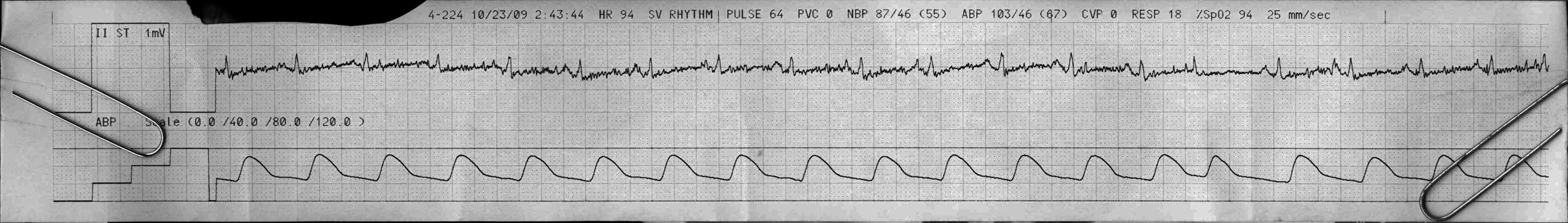
From across the room it looks like sinus.
Discussion:
Some smart cookie has cranked the gain on II up to 200%, and you can make out regular positive P waves and positive QRS complexes as expected in lead II. The voltage is really low, suggesting pathology or unusual patient positioning.
The P waves are almost as big as the QRS complexes, making me wonder about either weak ventricular forces, or big atria- maybe RAE.
There is a premature supraventricular beat near the end of the strip. It is probably junctional as we can’t see any prior P wave, but we can’t be sure it isn’t a PAC from just one lead.
Why does the monitor think this patient has a pulse of 64 bpm when they have over 90 QRS complexes per minute? Most likely this is a poor Sp02 tracing.
Final Impression?
Sinus rhythm @ ~90 bpm with one premature supraventricular beat.
Management implications:
12L to establish voltage and evaluate for chamber abnormalities. Check Sp02 waveform and adjust probe as necessary.
The Take-home Point:
A premature junctional beat does not follow a P wave, but it’s QRS morphology is generally identical to that of the sinus or ectopic atrial beats. Small changes in junctionally-paced QRS morphology can be caused by a superimposed retrograde P wave (modulating the QRS morphology). If the QRS starts to get wide, by definition it is from lower in the conduction system close to or beyond where the bundles branch off.
When differentiating narrow complex premature beats (PACs vs PJCs) note that not seeing a P wave, even on a clean baseline at adequate gain, does not mean there is no P wave! It just means there is no P wave seen in that lead. If the electrical forces are perpendicular to the axis of the lead, or are very well balanced, that lead will show no apparent activity. That’s why we look in more than one.
![]() One star strip. Students should identify the rhythm correctly.
One star strip. Students should identify the rhythm correctly.