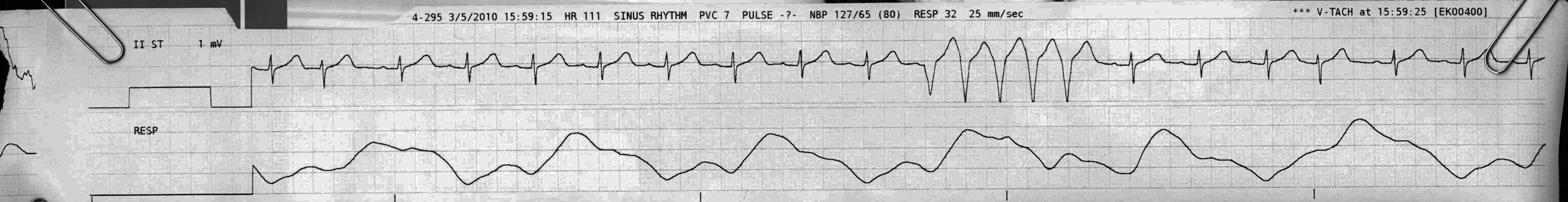
non-sustained VT
First Glance:
From across the room it looks like sinus rhythm with 5 beats of Ventricular Tachycardia (VT).
Discussion:
There’s not a lot to argue about in this one. It looks like VT. It smells like VT. So can you call it a run and get on with your life?
Probably. Although we have to mention that it’s hard to be 100% sure with only one lead. Sure, this looks perfect, but I have seen perfect-appearing pseudo-VT morphology that was movement artifact (and we saw an example in an earlier strip) so you must be careful to sniff around.
First, you can check the clinical correlation. Did the blood pressure drop? Did they become disoriented? That’s basic.
More interesting right now is retrospectively ruling out artifact. For this, you need to examine other leads. For instance, on this tele strip we only have lead II, but if you go to the machine you may be able to see the concurrent available limb and chest leads to see if they all display the same simultaneous changes consistent with VT.
While we’re on the subject, we can talk about superimposed waves in VT. Check this out:
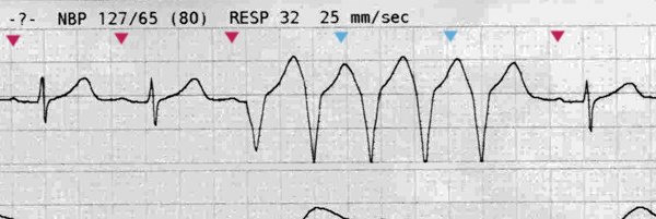
In magenta we see the atrial waves. Easy.
If this was pseudo-VT (artifact), we would probably see sharp deflections from the QRS waves superimposed on top of the artifact. We don’t see that here, which is a strong vote in favor of real VT.
However, in real VT you can often see the P waves superimposed on top of the wide ventricular QRS complexes, because in many hearts the AV node does not allow retro-depolarization from the idioventricular reentry. This evidence of AV dissociation (seeing regular P waves superimposed on a wide complex tachycardia) is pathognomonic for VT because it proves that the atria have nothing to do with what is going on and that the normal QRS complexes are not happening.
However, we don’t see them here because the P waves in this lead are diminutive. I know they’re there. The cycle does not appear to be reset and the magenta P wave after the run is exactly on time after two invisible blue P waves.
Final Impression?
Sinus rhythm ~90 bpm with non-sustained VT @ around 180 bpm with a 5-beat run.
Management implications:
All things being equal, PVCs, runs, and VT itself are all correlated with poorer electrophysiologic outcomes including sudden death. The monitor says this person has had 7 PVCs in the last minute, which is sufficient to hit the fairly arbitrary cut off for ‘significant’ ventricular ectopy already, and now you have a run.
The Take-home Point:
If it looks 100% like VT, fine, but be aware that there are lots of things that cause VT-like artifact. While this strip would satisfy me, anything less perfectly consistent with VT (especially if management would change based on the interpretation) would warrant evaluation of the simultaneous leads to confirm that this is not a lead-specific artifact.
![]() One star strip. Students should nail this.
One star strip. Students should nail this.