bigeminy
First Glance:
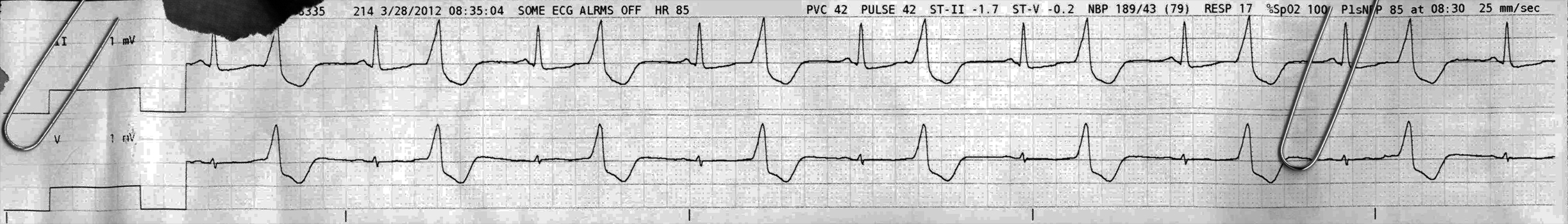
From across the room it looks like ventricular bigeminy.
Discussion:
We see here a regularly irregular rhythm. An apparently sinus beat is followed closely by a wide bizarre beat with no P wave. These couplets march out across the strip. This is ventricular bigeminy- where a PVC follows each sinus beat.
Here’s another strip from the same patient.
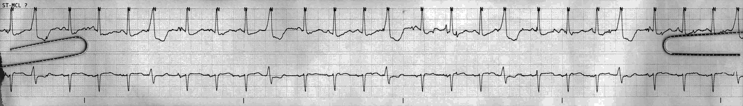
Now its ventricular quadrageminy. The upper trace is the same morphology because the limb leads look the same no matter what. The lower lead is very different, despite still being the ‘V’ lead. Most likely the V lead was placed in a very different spot since the prior strip.
You might be tempted to call P waves before many of the PVCs making you wonder about aberrant PACs, but I think that’s just movement artifact. Notice that they are all early, the interval is the same, and the morphology is identical to the prior strip with the clean baseline. The divots that look like they might be P’ waves are not always there, despite all this other regularity, and there are other random deviations. Those P’s waves must be artifact.
Note that the monitor, in its infinite wisdom, thinks that there are no PVCs per minute, and that every QRS is normal “N” as marked above.
One more strip:
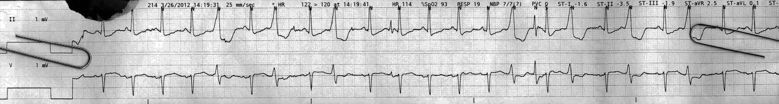
Here we see more of the same, now with irregular geminy. Sometimes couplets, sometimes more. We also see two narrow unusual QRS complexes (one small one before “HR 114” and one larger one under “PVC 0”). They don’t look like classic artifact. They aren’t in the right place for fusion beats. The subsequent beats, which follow very closely, conduct normally. They just don’t make any sense. Final verdict: artifact.
Final Impression?
Sinus rhythm with coupled PVCs, with a ventricular rate of ~85 bpm, and the latter two strips with a ventricular rate of ~ 110.
Management implications:
Bigeminy, as far as I can tell, is treated in the literature like a lot of PVCs. A lot of PVCs is worse than not-a-lot-of PVCs. The higher the ‘geminy’, the less irritable the ventricular myocardium probably is. The more PVCs you have, the higher the chance of significant arrhythmic problems, but the rhythm in and of itself is rarely a reason for cardiac compromise. It’s just a warning that it might get worse. After all- if you have excitable ventricles, such that they are firing every other beat, all they need to do is be a hair more excitable and you might have VT. If there are two or three firing simultaneously you probably have polymorphic VT. If there are a whole bunch of ventricular reentry circuits running at the same time, that is VF.
The Take-home Point:
‘Geminy’ is fun. It looks interesting. It’s usually a stable and perfusing rhythm. It’s quite common in the inpatient population and in my experience generally plays no part in management decisions. That does not mean you can ignore it, I’m just saying it is often ignored. Remember that increasing ectopy means the electrical milieu has changed and it is your job as the clinician to decide whether that is clinically relevant. Is the patient ischemic now? Lytes off? Too much albuterol? Who knows.
I regularly diagnose bigeminy from the physical exam. On exam the pt has a pulse (or PMI) where you can feel the couplet rhythm, but the second beat is weaker (presumably because it has had less diastolic filling time, and no atrial kick, and a ventricular contraction that was not perfectly coordinated by the purkinje system). If the patient has an S4 at baseline, you would not hear that with the PVC.
![]() Two star strip. The meat and potatoes.
Two star strip. The meat and potatoes.