wide complex tachycardia
First Glance:
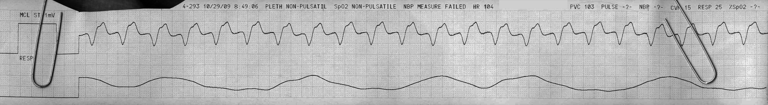
From across the room it looks a wide complex tachycardia.
Discussion:
Wide complex tachycardias, maybe the most devious yet clinically important differentiation problems for the non-electrophysiologist, is a long topic. We won’t be digging into it here, because you will not figure this out one from this one lead.
It’s clearly wide, with a QRS around 160ms. There is a deep QS wave. But this is MCL, which means without looking at the patient we have no idea what to expect. The very wide QRS makes me lean a little more towards VT, and if this is a septal lead it might push you towards LBBB-type VT because of the long delay to the nadir of the S wave here (see Brugada criteria for wide complex tachycardia differentiation).
The rate is on the slow side for VT, and on the fast side for an accelerated idioventricular rhythm (which is also ventricular, but might be an accelerated escape rhythm instead of a reentry pathway as in classic VT). If we could see P waves that might push us towards sinus or VT (depending on where they are) but they aren’t obvious here.
We see ST elevation, but it’s significance is completely unclear given we may have LBBB here.
Final Impression?
Wide complex tachycardia @ ~110 bpm.
Management implications:
Figure this out! In the stable patient a 12L will be extremely helpful.
The Take-home Point:
You generally aren’t going to feel comfortable calling VT from a single strip unless you see AV dissociation, or know the pt has a normal EKG and/or it is a witnessed/paroxysmal finding.
If someone comes in with palpitations but otherwise fine, you put them on the monitor and see this, and you have no prior EKGs- the only way I will be able to call VT from the one lead is if I see AV dissociation. Ie: If you can clearly see regular asynchronous P waves superimposed over the rhythm. We don’t see that here.
![]() Two star strip. The meat and potatoes.
Two star strip. The meat and potatoes.