sinus rhythm with artifact

First Glance:
From across the room it looks like a low voltage sinus rhythm with fuzzy artifact.
Discussion:
Again with the artifact. We can see the arterial pulse marching on so the fuzz on the lead II trace is clearly artifact, and it’s certainly too fine/high-frequency to be any kind of coordinated electrical activity of any chamber. In my experience, this kind of artifact is most likely caused by muscle use. For example, we see the artifact in lead II, so perhaps the patient exerted their right arm or left leg with a distal electrode.
Look at the arterial trace near the end of the strip. Can you pick out the PAC?
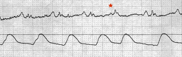
The P waves are regular and identical across the strip until the starred beat, which is smaller. Notice that the a-line trace shows the same systolic pressure achieved after only 460ms (this would be a heartrate of 130 bpm). This is reasonable, because we are not getting into the higher rate tachycardias where reduced preload in a premature beat result in reduced cardiac output.
How about those QRS complexes? They are smaller than the P waves in lead II. This could be low-voltage, or it could be atrial enlargement, or it could be nothing.
Final Impression?
Sinus rhythm @ ~90 bpm.
Management implications:
Nothing special. 12L if you are worried about low voltage and flat T waves.
The Take-home Point:
Don’t call ‘low-voltage’ from a tele strip, especially not just one lead. There are a variety of ways to get this, including nonstandard positioning for electrocardiography (ie: sitting upright, etc).
![]() One star strip. Students should identify the rhythm correctly.
One star strip. Students should identify the rhythm correctly.