sinus rhythm
First Glance:
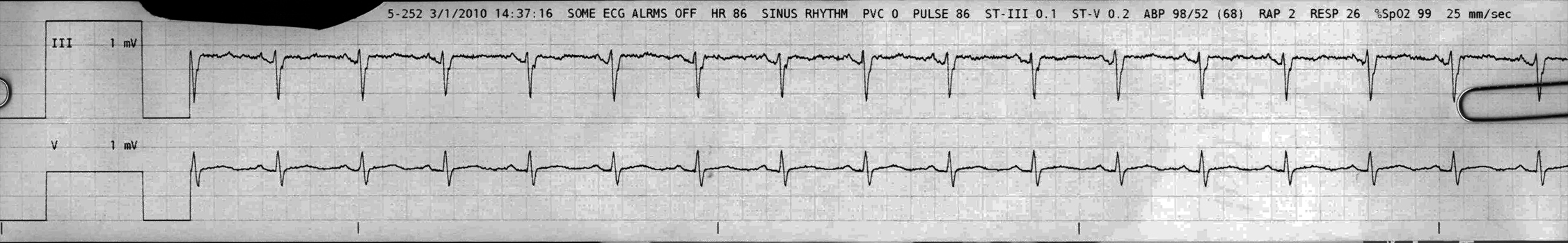
From across the room it looks like sinus rhythm.
Discussion:
This looks like sinus rhythm, with a positive P in lead III (as expected) with flattened inverted T waves. The QRS complexes are narrow, with a hint of rSr’ in III which is not unusual and clinically unremarkable. Sometimes when I see that ‘divot’ in the terminal part of the lead III QRS I wonder whether there is a superimposed P wave (these complexes look like many junctional or reentry rhythms with a buried retrograde P), but here that is not the case because the P waves are up front where they are supposed to be.
Where is the V lead placed? Usually folks place it smack in the middle which would show the septal (v1, v2) area.
The V lead has a positive P with a slightly positive net deflection, which would be normal around V3 or 4 in most patients. Also note that the T wave is upright in V which would not be expected in the septal region.
If you look closely it looks like there is PR segment depression in both leads, more obvious in III where the voltage gain is at 200%. This could be seen in pericarditis (although we don’t see concurrent ST elevations in either of these leads) or in atrial ischemia. It’s a subtle finding, however, likely nothing (possibly some artifact of the tele trace processing) and other leads would need to be evaluated.
Final Impression?
Sinus rhythm @ ~85 bpm, possibly with nonspecific atrial repolarization abnormalities.
Management implications:
Maybe a 12L to evaluate PR segments, or maybe not.
The Take-home Point:
PR segment depression is a pathologic finding. The true ‘baseline’ of the EKG is the flat segment before the P wave, not after it, and I understand that PR depression and ST elevation should both be compared to the flat terminal QP segment as a reference.
![]() One star strip. Students should identify the rhythm correctly.
One star strip. Students should identify the rhythm correctly.