sinus rhythm with PACs

First Glance:
From across the room it looks like sinus with some supraventricular ectopy.
Discussion:
The atrial activity is seen better in the lower ‘V’ (chest) lead. It’s normal sinus rhythm, but then at beats 2, 7, and the last beat we see PACs.
Here is one:
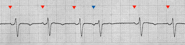
We can see that these PACs are preceded by a P wave that is early (premature), has a normal PR interval (atrial), and results in a depolarization of the ventricle through the normal conduction tracts (contraction). PAC = premature atrial contraction. We can see that the morphology of this early P wave is different (no positive deflection), consistent with an ectopic beat.
After the PAC there is a pause (a little longer than a non-compensatory cycle) and the sinus rhythm resumes.
The positive then negative deflections in the P wave of the chest lead are consistent with a septal lead, where T inversion is normal. Lead II looks remarkable only for a flattened T wave.
Final Impression?
Sinus rhythm @ 85 bpm with three PACs
Management implications:
Ectopy can be a sign of irritated myocardium or a hyperactive sympathetic system. PACs don’t cause hemodynamic problems, however, especially not at these calm rates.
The Take-home Point:
The PR interval of this PAC is normal.
If the PR interval is normal, then the P wave originated in the atria. If the PR interval is short, then either it originated in the atria but was conducted unusually rapidly (rare), there is an accessory pathway (WPW, etc), or it originated below the atria (AV node or below).
![]() One star strip. Students should identify the rhythm correctly.
One star strip. Students should identify the rhythm correctly.