SSS vs nonconducted atrial ectopy
First Glance:
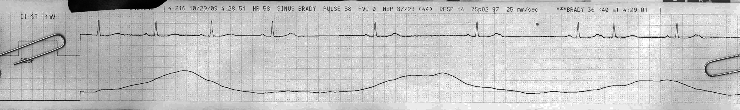
From across the room it looks like an irregularly irregular supraventricular bradycardia.
Discussion:
Grossly, across the first four beats, it looks like sinus rhythm @ ~60 bpm, followed by a lost P wave. One way to get that kind of strip would be a sinoatrial block, but the next beat is nowhere near where it would be expected if we had just dropped one sinus pace.
So maybe it’s sinus pause/arrest? That is a possibility. But look at the T waves:

I see that the first three T’s are identical with smooth positive monophasic deflections, then beat 4 has a bifid T with a sharp terminal positive deflection (orange triangle). This is the native T with a P wave slapped on top- although it’s unclear whether it’s a sinus P or an ectopic atrial depolarization.
We see these stacked T/P’s at beat 4, 5, 6 and 7 and on the 7th beat it actually conducts (albeit with a longer PR interval) and there is a subsequent QRS. That could be a sinus beat, or a PAC. A long strip might help differentiate (if we caught some atrial ectopy that wasn’t superimposed). The longer PR interval on that conducted beat might suggest some concurrent AV node pathology (ie: tendency to Wenckebach).
A variety of sinus abnormalities could cause this, and none of them are benign. Abnormal and unpredictable sinus activity is consistent with ‘sick sinus syndrome’, which is a broad term for a variety of dysfunctional sinus problems.
Another good possibility is that these are nonconducted PACs. If those superimposed P waves are premature atrial ectopy, but in the refractory period of the AV node or purkinje fibers, then they won’t conduct down the fibers and fire the ventricles. However, we would have expected the next sinus P after this ectopic ‘reset’ to come earlier- probably about one R-R interval after the ectopic beat. Here they are distinctly longer than that.
Of note is that despite long pauses (up to just under 2 seconds in this strip) there is no escape beat. We would expect a junctional escape beat well before the next sinus beat comes in, which means we can’t rely on the AV node to back up the crippled sinoatrial pacemaker. This is further evidence for disease of the AV node, along with that extended PR interval in the conducted couplet discussed earlier.
Final Impression?
Sick sinus syndrome vs sinus bradycardia with nonconducted PACs, with pauses to 2 seconds, for a ventricular rate of 45.
Management implications:
With a ventricular rate of 45 over the course of this strip, no reliable escape rhythm, the monitor probably ringing off the hook about the pauses, and a last BP measured with a MAP of 44, we will pay attention to this bradycardia. If sustained it would likely be symptomatic. For the time being monitoring and supportive care will be important.
As this bradycardia does not appear to be caused by high grade AV blocking, it would be more likely to respond to atropine in an acute situation.
The Take-home Point:
The dire differentials suggested above are the most likely ones in an ill hypotensive patient admitted on tele. However, another reasonable differential for sinus bradycardia plus suppressed junctional escape and the apparent beginning of a 2nd degree type I sequence (last couplet) could be a patient with extremely high vagal tone. If this strip was off of a healthy 24yo cross country runner while sleeping, during an admission for an elective ortho procedure with excellent pain control, you might come up with the exact same strip but with no management or prognostic implications whatsoever. Although to resolve that arrhythmia all you should need to do is wake him up and have him get his heart rate up.
![]() Three star strip. Devious stuff.
Three star strip. Devious stuff.