atrial fibrillation
First Glance:
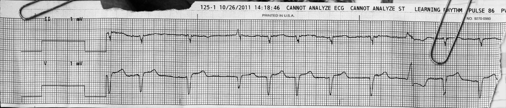
From across the room it looks like atrial fibrillation with a PVC.
Discussion:
The fibrillatory baseline is consistent with afib, supporting that diagnosis in the setting of yet another irregularly irregular rhythm. There are a couple unusual QRS complexes.
The fourth beat looks like a fusion beat, with characteristics of the native QRS (best seen in the lower lead) and a fairly normal ST / T wave, but the axis and deflection amplitudes are different.
There is a PVC three beats from the right.
The lower lead ‘V’ is a chest lead, with one millimeter of ST elevation. We can see it is at 50% voltage calibration, so this would be 2mm if gained up to standard calibration.
Here’s another strip from the same patient:
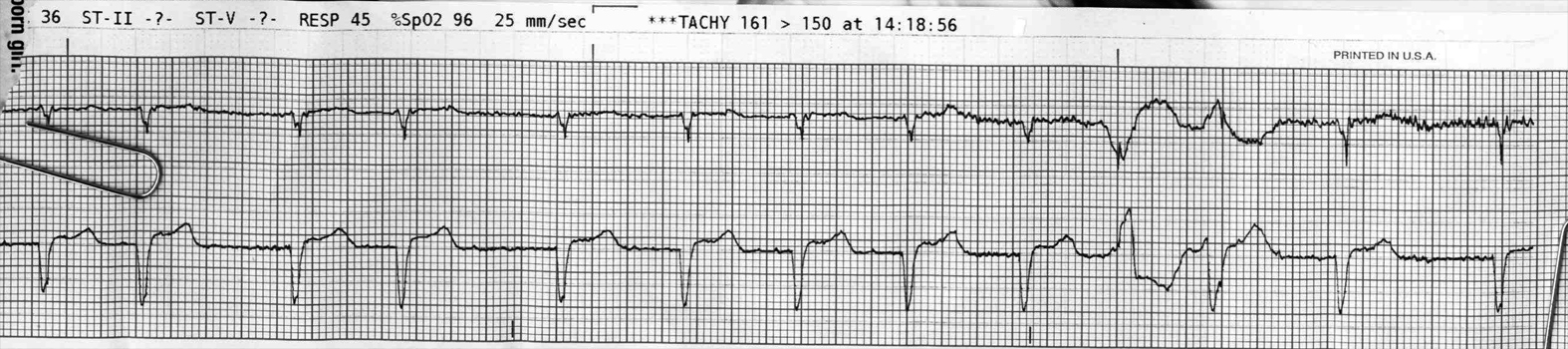
There are extremely sharp negative spikes associated with all QRS’s in the upper lead, that look like pacing spikes (purple triangles below). However, their appearance in this strip is inconsistent with any functioning pacing device, and there is no biological/endogenous way to generate spikes of such rapid/narrow deflection. These are likely artifact of some type, although I am open to suggestions..
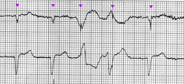
It looks like there is some multifocal ventricular ectopy at beat 3 and 4 of this excerpt, or maybe a PVC and then a fusion beat, but I don’t actually think these are PVCs. I think most of what we see is movement artifact.
Beat 3 looks like a PVC, with a wide complex and appropriate discordance, but if you assume the steep downwards deflection is the same thing we see in the native QRS complexes, then followed by some movement artifact, we can see the peak of the T wave at the expected interval. Beat 4 looks more like the native QRS- maybe with a little aberrancy or some movement artifact.
Final Impression?
Atrial fibrillation @ ~90 bpm, with ST elevation in the chest lead.
Management implications:
12L to evaluate for ischemia.
The Take-home Point:
A fusion beat is a fusion of a supraventricular beat and a ventricular beat. It generally has characteristics of both the native supraventricular QRS but is wider, or somehow aberrant. For example: if a patient is having monomorphic ventricular ectopy, the fusion beat will often look like a hybrid between the native QRS and the PVCs.
![]() One star strip. Students should identify the rhythm correctly.
One star strip. Students should identify the rhythm correctly.