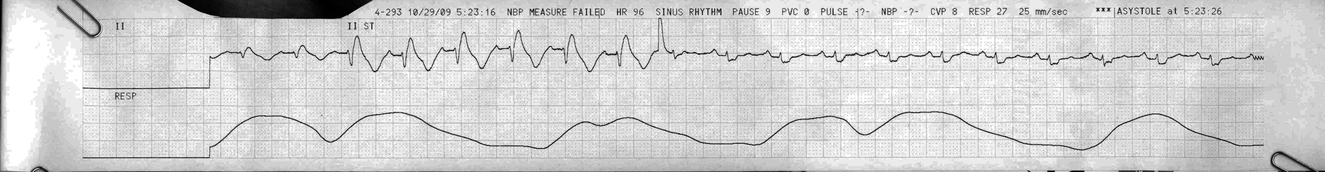
Sinus tach, and messing-around-with-the-monitor artifact
First Glance:
From across the room it looks like a series of wide-complex beats.
Discussion:
Look closely. We start in lead II with unknown voltage calibration and see that on beat three the QRS gets big and exciting, but that the morphology is identical to the first two beats. After beat 8 the strip abruptly changes again, but this time to a QRS with a completely different morphology/axis.
Is this two distinct rhythm changes in 15 seconds?
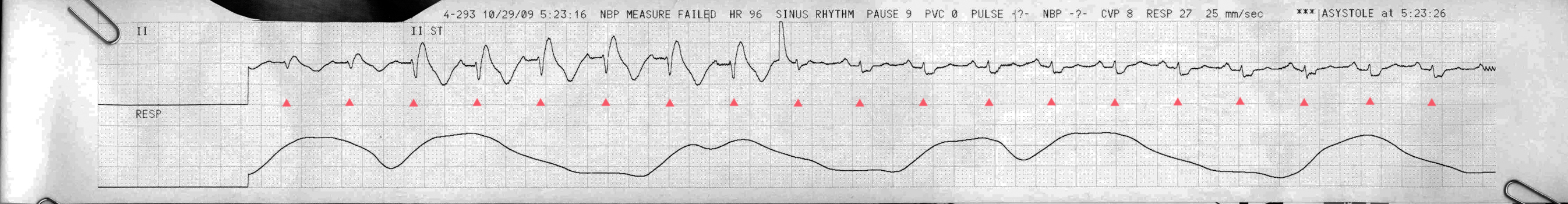
Nope. We can easily track out the rhythm which maintains its regularity across the strip, and we can see regular P waves before every QRS.
This is probably someone at the bedside increasing the gain on the voltage calibration (first change, where the gain goes up) and then the lead (second change, where the gain goes down and the morphology/axis changes). That blip when it changes the second time is not a PVC- it’s the monitor re-zeroing as it switches to a new lead.
So what is the rhythm then? It looks like a borderline sinus tachycardia, with a long PR interval, and a wide complex with ST segment changes (ST elevation and T inversion in the early complexes, and exactly opposite that in the later ones). The morphology is weird, especially as it is hard to see in the early complexes where the QRS ends and the ST begins, making it possible that it is not a QRS with significant ST elevation, but rather a wide and bizarre complex that looks like that seen in hyperkalemia or an agonal rhythm or TCA overdose. However, it’s probably none of those latter options because the P is unusually well defined for either of those clinical scenarios, in my very limited experience.
The person evaluating this patient was probably just as curious as we originally were, hence increasing the gain to reveal the P waves better (though it was still hard to tell whether they were P waves, or the tail end of an unusual QRS). Then they switch leads to find a better look at the atrial activity.
Final Impression?
Wide complex sinus tachycardia at ~100 bpm, borderline first degree block, with ischemic changes.
Management implications:
Compare to prior morphology, check labs (?K), and get another 12L.
The Take-home Point:
We generally look to lead II and V1 for most rhythm assessments as they tend to reveal the atrial activity best. However, sometimes another lead will provide a better (or complimentary) look, and changing the gain or paper speed on the monitor or the 12L machine can give you the data you need to call it.
![]() One star strip. Students should identify the rhythm correctly.
One star strip. Students should identify the rhythm correctly.