atrial fibrillation
First Glance:
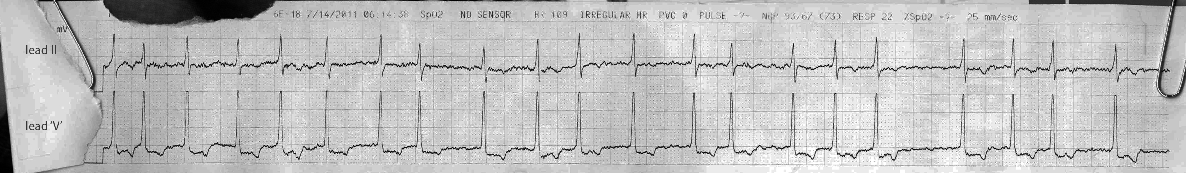
From across the room it looks like afib.
Discussion:
The rhythm is clearly irregularly irregular, and I can see nothing with any hint of an organized atrial rhythm. The ventricular rate is 109 according to the monitor. This looks like afib with Rapid Ventricular Response (RVR).
In both leads we see that there are inverted T waves, which is concerning, as well as a bit of ST depression in the V lead. This is common in ill patients where the perfusion demands of a tachycardia could cause some low grade cardiac ischemia (‘demand ischemia’), or it could be an AMI, or some other nonspecific repolarization change.
A student I shared this with wondered whether there was a hint of Left Ventricular Hypertrophy (LVH) in this strip with the tall R waves in ‘V’ and the possible strain pattern in this one lateral lead, but you can’t make that call for two reasons. First, and more importantly, you have no idea which precordial lead this is. The lead marked ‘V’ on the 5-lead monitor that I see in most hospitals can be put anywhere. It can be placed at V1 or V2 and give you a right-ventricle-predominant view, or it could be placed further lateral and give you an LV predominant view, so this tall R wave could be Right Ventricular Hypertrophy (RVH) in the former, or LVH around V4, or nothing in V5/V6 (because it does not meet LVH voltage criteria). Second, and less importantly, you have no idea what the calibration is from this strip because it was folded over. I happen to know it’s at 120% for the V lead, which isn’t helpful either.
Knowing where the V lead is would really be helpful here. If it was in V1 or V2 we should pay attention. Tall R waves with ST depression in the septal region could be right ventricular hypertrophy or strain, or posterior MI with ‘reversed’ Q waves and ST elevation.
Final Impression?
Afib with RVR at 110 bpm, suspicious for nonspecific ischemia.
Management implications:
Check lead placements. Evaluate for ischemia with 12L. Can consider getting additional posterior (V6, 7, and 8) leads with the 12L. Deal with afib rate control per usual.
The Take-home Point:
Limb leads, as long as they are placed in the correct order, don’t care much where they are. You can put the right arm lead (for instance) on the right hand, the wrist, the elbow, the shoulder, or even further proximally and get a clinically similar (or often identical) series of tracings.
Chest leads, however, are supremely reliant on positioning and it takes far more skill and patience to place them correctly in the standard positions. When commenting on critical findings in a precordial/chest/’V’ lead- be sure you know where it is reading from!
![]() One star strip. Students should nail this.
One star strip. Students should nail this.
Great opening case! Good because it shows a common arrhythmia with some equally common additional pathology. I particularly liked the discussion section where it expanded on the likely pathology and commented on the implications of lead placement. At my workplace I tend to only get one strip from one lead, usually lead II, but good to think about chest leads as well. The structure of the discussion is good as well in terms of the headings. Has definitely made me want to go through more cases and subscribe!
Pingback: The ECG Resources I use | smacem